What does mind control look like? Visualisation of parasites in hosts

Researchers from the Natural History Museum together with colleagues from UK and Canada are developing new innovative techniques to visualise the interactions between liver fluke parasites and their ant hosts. Improving the understanding of these interactions can serve as a model for future studies to visualise host-parasite interactions in the intermediate hosts of NTDs. NHM’s Dr Martin Hall and Dr Daniel Martin-Vega explain more.
The small liver fluke, Dicrocoelium dendriticum, is a trematode parasite that infects wild and domestic mammals and very occasionally humans. To reach its mammal hosts its complex life cycle relies on first infecting snails and then ants which are ingested by a grazing animal such as a deer or a cow.
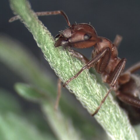
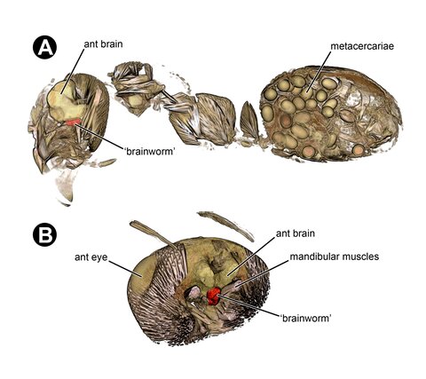
In the intermediate ant host, most D. dendriticum individuals become infective larvae (metacercariae) lodged in the ant’s abdomen waiting for the ant to be eaten by a passing mammal. Rather than just leaving this to chance, one D. dendriticum parasite (or rarely more than one) will lodge itself into the ant’s brain and begin to take control, inducing in the ant an atypical, suicidal behaviour. When the environmental conditions are right the parasite causes the infected ant to climb to the top of grasses and other low-level vegetation to which they cling with their mandibles. Once in this position the ants are much more likely to be ingested by grazing ruminants, thereby enabling the flatworms to infect their new hosts in the livers of which they develop into adults.

Traditional techniques used to image the parasites inside the ants are particularly challenging due to the ants’ hard cuticle and fragile brain tissues. However, X-ray micro-computed tomography facilitates a non-destructive, virtual histological dissection at any slice orientation to reveal the 3D structure of biological objects. Here we stained infected ants with phosphotungstic acid (PTA) and then scanned their decapitated heads and separated abdomens using a ZEISS Xradia 520 Versa.
Our study demonstrates that micro-CT is a promising tool for providing unparalleled novel insights into the physical interface between parasites and their hosts. For the first time we were able to show the infestation of an ant head not only with the, so-called, ‘brain worm’, but with metacercariae which, up until now, had only been illustrated in the abdomen.
We also show infestation of the brain with up to three brain worms, provide novel qualitative information on the location of infestations and quantitative data on the parasites themselves, including volumetric data for comparisons between the different parasite forms. The results from the present project were presented at the 26th International Conference of the World Association for the Advancement of Veterinary Parasitology (Kuala Lumpur, Malaysia, 4th-8th September 2017) and are being prepared for publication.